Medical imaging: what you need to know
Published 22 September 2022

© Crown copyright 2022
This publication is licensed under the terms of the Open Government Licence v3.0 except where otherwise stated. To view this licence, visit nationalarchives.gov.uk/doc/open-government-licence/version/3 or write to the Information Policy Team, The National Archives, Kew, London TW9 4DU, or email: psi@nationalarchives.gov.uk.
Where we have identified any third party copyright information you will need to obtain permission from the copyright holders concerned.
This publication is available at https://www.gov.uk/government/publications/medical-imaging-what-you-need-to-know/medical-imaging-what-you-need-to-know--2
One of the biggest advances in modern medicine has been the use of medical imaging to help diagnose and treat patients. Most people will have some form of medical imaging in their lifetime. Common examples are:
- X-rays
- CT (computed tomography) scanning
- ultrasound
- MRI (magnetic resonance imaging)
- nuclear medicine imaging
Some medical imaging uses radiation. This guide will tell you about the benefits of different types of medical imaging as well as the radiation risks.
There are significant benefits to you from getting the right diagnosis and treatment. To make sure you get the right examination your healthcare professional will ask you about your medical history, explain the options available and discuss what is most important to you.
If medical imaging is needed, they will send a request to an imaging specialist. This specialist will check the request and make sure that you get the best imaging examination for you. Imaging examinations are not always needed and so in some cases, you may proceed straight to treatment.
This guide gives you some information on the different types of imaging examinations and their benefits and risks. Your healthcare professional will make sure that the benefits to you personally of having any examination are greater than the radiation risks. You can use the table at the end of this guide to look at the radiation risks for different examinations.
If you still have questions after reading this guide, please ask your healthcare professional.
X-rays
Almost as soon as X-rays were discovered they were used in medical imaging. An X-ray machine is used to produce radiation which passes through your body to create an image. Different parts of your body absorb the X-rays in different amounts. Soft tissue and lungs absorb less X-rays than teeth and bones, so they look darker on X-ray images. Teeth and bones look whiter on the images.
Some examples of how X-rays are used
Dental radiography
This is the most common type of X-ray examination. Millions of dental X-rays are taken every year in the UK. Dental X-rays are used to diagnose disease in the mouth such as, tooth decay or gum disease. They are also used to plan treatments and monitor the health of your teeth.
General radiography
Uses X-rays to produce 2D images of the inside of your body. X-ray examinations help to diagnose conditions such as chest infections and look for broken or damaged bones. X-rays are also used in scans to monitor bone density (DXA).
Fluoroscopy
Uses X-rays to produce moving images of parts of your body which can be displayed on a screen. This is used to diagnose and monitor conditions such as your digestive system, blood flow (cardiovascular system) and urinary system. Depending on the type of examination, a dye that shows up on X-ray might be used, this may be injected or swallowed.
Interventional radiology
Uses X-rays to produce moving images which can be used to guide and deliver treatment. It is used to diagnose and treat a broad range of conditions such as blockages in your heart, blood vessels, food pipe (oesophagus) and kidneys. It is also used to guide doctors to an area of disease to take a sample (biopsy) or deliver treatment. This type of imaging and treatment can reduce the need for surgery.
Computed tomography (CT)
Uses X-rays to produce 3D images of your body. The images are used to diagnose, guide treatment and monitor a broad range of diseases and injuries. Some CT scans require you to have a small tube put into your arm so that dye that shows up on X-ray can be injected during the examination. You may also be asked to drink water or a mixture of water and dye before your scan. These will help different parts of the body to be seen more clearly.
Nuclear medicine
Uses a small amount of a radioactive substance that is usually injected into a vein (or it is swallowed or inhaled). The scanner will detect this low-level radioactivity coming out of your body and will build up images. These images show how your body is functioning. They are used to diagnose, aid treatment and monitor a wide range of diseases and conditions. The radioactivity in your body usually falls to an undetectable level in a few days.
PET-CT or PET-MRI scanning
PET (positron emission tomography) is a type of nuclear medicine scan that produces 3D images in combination with CT or MRI. A small amount of a radioactive substance is injected into a vein. PET images are used to diagnose and monitor a wide range of diseases and conditions. The radioactivity in your body usually falls to undetectable within a day.
Ultrasound and MRI
These are types of imaging that do not use X-rays or radioactive substances. They can be used to diagnose a range of conditions but cannot be used for everything. The imaging specialist will always consider these types of imaging if they are an option for you.
Pregnancy and breastfeeding
For some examinations we will need to know if you may be pregnant or breastfeeding. This is to make sure that any radiation risk to the foetus or baby is kept as low as possible.
We don’t always need to ask. It will depend on the examination you are having if you are asked whether you are pregnant or breastfeeding.
When the benefit to you clearly outweighs the small risk from the radiation the examination may go ahead after discussing all the options with you. The risk of not carrying out the examination can be much greater than the small risk from radiation.
If you are breastfeeding and need to have a nuclear medicine examination, you may need to stop feeding for a while, or stop altogether. You will be given advice and guidance on any precautions that you need to take.
Risks
Everything we do in our daily lives carries some level of risk. We tend to regard activities as being ‘safe’ when the risk of something unpleasant or unwanted happening falls below a certain level. The lower the level of risk, the ‘safer’ the activity is seen to be.
People judge risk on both how likely an event is and how we would feel if it happened. That means everyone has a different perception of the same risk, because it depends how they feel about the event happening.
Every type of medical imaging examination carries with it some risks and some benefits. Your healthcare professional can help you to understand how this applies to you.
Effects of radiation
We know from studying people who have been exposed to high doses of radiation that this can increase their chances of developing cancer much later in life. However, the amounts of radiation used in medical imaging are very small and have very low risks.
Any exposure to radiation from medical imaging is kept as low as possible to reduce the risks. Each time you are offered an examination you and your healthcare professional will weigh up the risk and benefits to you.
Natural risks
We know many people develop cancer, in fact about 1 in 2 (50%) of us will get cancer in our lifetime. The chances of any of us developing cancer are affected by all sorts of things, such as our genes, exposure to smoke, diet, weight, and alcohol intake. We have tried to estimate how much the radiation from each examination will increase your chances of developing cancer.
The figure below shows how even the highest dose examinations would only increase this risk from around 50% to 51%, a tiny increase considering the benefits to you from having the examination. The most common examinations would only increase this risk by much less than 1%.
Figure 1. Diagram of the typical levels of risk associated with each type of examination.
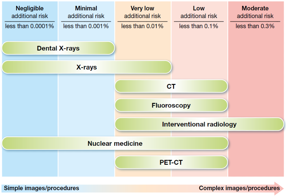
This figure is divided into 5 columns, each indicating an increasing amount of risk, and then shows how these risks relate to different imaging procedures. The 5 columns are:
- negligible additional risk – less than 0.0001% risk
- minimal additional risk – less than 0.001%
- very low additional risk – less than 0.01%
- low additional risk – less than 0.1%
- moderate additional risk – less than 0.3%
The procedures shown on the diagram are:
- dental X-rays – negligible to minimal risk
- X-rays – negligible to very low risk
- CT – very low to low risk
- fluoroscopy – very low to low risk
- interventional radiography – very low to moderate
- nuclear medicine – negligible to low risk
- PET-CT – very low to low risk
This guide had been produced by UKHSA and supported by: Society of Radiographers and College of Radiographers, British Dental Association and Royal College of Radiologists.